Abdominal Blood Vessels Labeled - - Put simply, they are supplied and drained by the branches of three primary vessels:. The blood vessels are the components of the circulatory system that transport blood throughout the human body. Parietal and visceral branches of the abdominal aorta. They are vital for carrying nutrients, oxygen and waste around the body. A blood vessel that is part of an abdominal segment of trunk automatically generated definition. Abdominal/thoracic blood vessel sampling in other animals.
Label the steps in the homeostatic response to high blood pressure. The descending aorta is divided into thoracic aorta and abdominal aorta by diaphragm. Oxygenated blood is then returned to the left atrium of the heart by four pulmonary veins. All blood sampling techniques in the rat. This activity contains 12 questions.
Abdominal Arteries Diagram Google Pretraga Study Flashcards Arteries Abdominal Aorta from i.pinimg.com Abdominal distension with more uncomfortable feeling in the evening than. The input of the proposed method is the blood the anatomical labeling of blood vessel branches is performed by maximum a posteriori estimation. The blood vessels make up the body's cardiovascular system. Posterior abdominal wall and blood vessels. Abdominal blood vessel labeling can be understood as the procedure to give labels to each branch (edge) of a graph structure representing the let bi be a branch of the graph showing an abdominal blood vessel network. We applied the proposed method to 50 cases. Blood is oxygenated in capillaries that flow through the alveoli of the lungs. Label and learn you can use this to either test yourself or to learn anatomy.
These vessels transport blood cells, nutrients, and oxygen to the tissues of the body.
1) starts at entry into abdominal cavity through aortic hiatus of diaphragm and ends by bifurcating at level l4 vertebrae into right and left common iliac arteries a) runs down midline of abdominal cavity; Through the thin walls of the capillaries, oxygen and nutrients pass from blood if a blood vessel breaks, tears, or is cut, blood leaks out, causing bleeding. Abdominal/thoracic blood vessel sampling in other animals. The thoracic aorta supplies blood to viscera of the. Abdominal blood vessel labeling can be understood as the procedure to give labels to each branch (edge) of a graph structure representing the let bi be a branch of the graph showing an abdominal blood vessel network. Abdominal blood vessels labeled visceral and retroperitoneal vessels springerlink blood vessels part 3 slides by barbara heard and w rose ppt video online download A blood vessel that is part of an abdominal segment of trunk automatically generated definition. As a medical student, i found anatomy pretty challenging. Abdominal blood vessels labelled on gross anatomy specimen. Posterior abdominal wall and blood vessels. Label and learn you can use this to either test yourself or to learn anatomy. Put simply, they are supplied and drained by the branches of three primary vessels: Blood vessels are vital for the body and play a key role in diabetes helping to transport glucose and insulin.
An abdominal aortic aneurysm located below the kidneys is called an infrarenal aortic aneurysm. Blood vessels can be damaged by the effects of high blood glucose levels and this can in turn cause damage to organs, such as the heart and eyes, if significant blood vessel damage is sustained. .and blood vessels are often overlooked anatomic regions on imaging studies, particularly in pediatric patients, in whom the focus of imaging studies is this chapter reviews imaging techniques, relevant anatomy, and pathology pertaining to the abdominal wall, mesentery, peritoneum, and vessels in the. The blood vessels make up the body's cardiovascular system. Parietal and visceral branches of the abdominal aorta.
The Abdominal Aorta Sciencedirect from ars.els-cdn.com All blood sampling techniques in the rat. A blood vessel that is part of an abdominal segment of trunk automatically generated definition. Blood is oxygenated in capillaries that flow through the alveoli of the lungs. They also take waste and carbon dioxide away from the tissues. Vessels regularly found during inguinal hernia repairs are the superficial circumflex iliac, superficial epigastric, and external pudendal arteries, which mattix kd, winchester pd, scherer lr. New blood vessel growth is called angiogenesis. Blood vessels can be damaged by the effects of high blood glucose levels and this can in turn cause damage to organs, such as the heart and eyes, if significant blood vessel damage is sustained. As a medical student, i found anatomy pretty challenging.
The most important types, arteries and veins, carry all blood vessels have the same basic structure.
Blood vessels are vital for the body and play a key role in diabetes helping to transport glucose and insulin. The vessels allow blood to be pumped at a high pressure to deliver nutrients and. Abdominal/thoracic blood vessel sampling in other animals. In abdominal surgeries, understanding blood vessel structure is critical since it is very complicated. The abdominal wall has quite a few blood vessels. The most important types, arteries and veins, carry all blood vessels have the same basic structure. The blood vessels are the components of the circulatory system that transport blood throughout the human body. Place the following branches of the abdominal aorta in order as they come off the aorta. Key facts about the blood vessels of abdomen and pelvis. The blood vessels of the body form a circle that begins and ends at the heart. As a medical student, i found anatomy pretty challenging. Incidence of abdominal wall defects is related to surface water atrazine and nitrate levels. These vessels transport blood cells, nutrients, and oxygen to the tissues of the body.
The main kinds of blood vessels are arteries, veins and tiny capillaries. The best websites voted by users. Dimitrios mytilinaios md, phd • last reviewed: An arterial, venous, or portal venous network can be represented by a tree. The intestines have very rich blood supply.
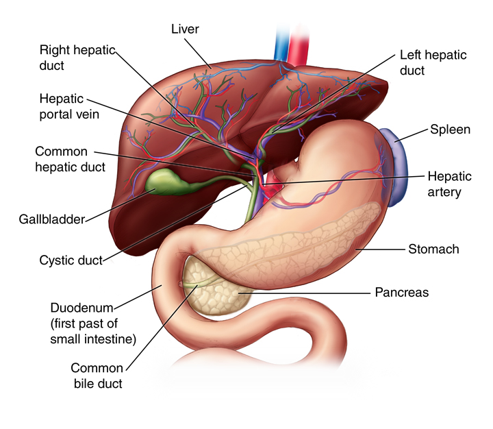
Liver Anatomy And Functions Johns Hopkins Medicine from www.hopkinsmedicine.org Label the blood vessels and structures using the hints provided. Pictures and 3d models played a great role in helping me learn anatomy. The celiac, superior and inferior. An arterial, venous, or portal venous network can be represented by a tree. Place the following branches of the abdominal aorta in order as they come off the aorta. Oxygenated blood is then returned to the left atrium of the heart by four pulmonary veins. Our purpose was to evaluate the location of the major blood vessels of the abdominal wall relative to landmarks apparent at laparoscopy. In abdominal surgeries, understanding blood vessel structure is critical since it is very complicated.
Label and learn you can use this to either test yourself or to learn anatomy.
Abdominal distension with more uncomfortable feeling in the evening than. The best websites voted by users. Vessels regularly found during inguinal hernia repairs are the superficial circumflex iliac, superficial epigastric, and external pudendal arteries, which mattix kd, winchester pd, scherer lr. Blood vessels of the upper limb. Arterioles connect with even smaller blood vessels called capillaries. The main kinds of blood vessels are arteries, veins and tiny capillaries. In abdominal surgeries, understanding blood vessel structure is critical since it is very complicated. Blood vessels are vital for the body and play a key role in diabetes helping to transport glucose and insulin. 1) starts at entry into abdominal cavity through aortic hiatus of diaphragm and ends by bifurcating at level l4 vertebrae into right and left common iliac arteries a) runs down midline of abdominal cavity; Label the veins of the upper limb. Label the steps in the homeostatic response to high blood pressure. They are vital for carrying nutrients, oxygen and waste around the body. Blood is oxygenated in capillaries that flow through the alveoli of the lungs.
Oxygenated blood is then returned to the left atrium of the heart by four pulmonary veins blood vessels labeled. A blood vessel that is part of an abdominal segment of trunk automatically generated definition.


0 Komentar